
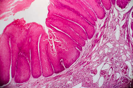
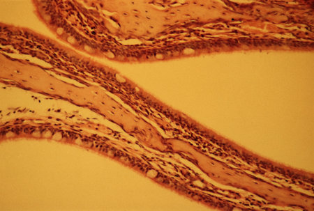
Condyloma acuminatum, also known as genital warts. Light micrograph, photo under microscope
Коллекция по умолчанию
Коллекция по умолчанию
Создать новую

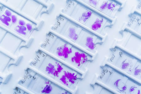
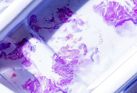
Rows of microscope glass slide in the cells
Коллекция по умолчанию
Коллекция по умолчанию
Создать новую

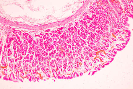
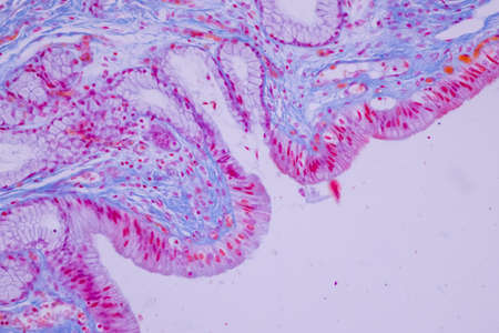
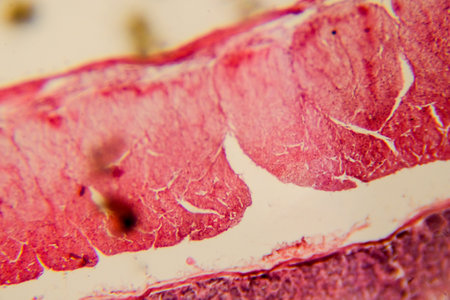
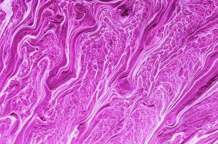
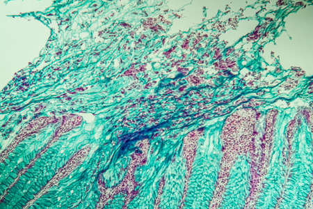
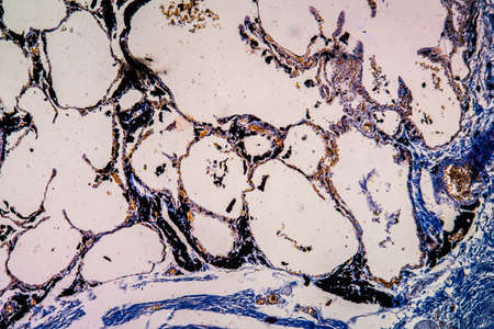
Abstract science background- pyloric division of the stomach of the dog. Cell biology
Коллекция по умолчанию
Коллекция по умолчанию
Создать новую

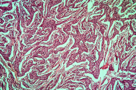
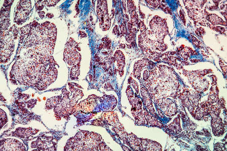
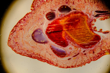
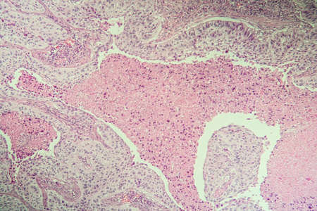
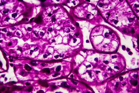
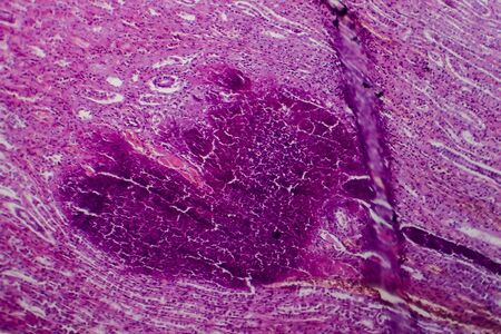
Ovarian cancer, light micrograph, photo under microscope. Photograph shows a fragment of a cancerous tumor in the female ovary. Selective focus
Коллекция по умолчанию
Коллекция по умолчанию
Создать новую
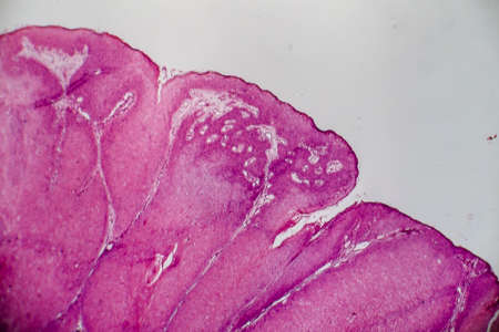
Condyloma acuminatum, also known as genital warts. Light micrograph, photo under microscope
Коллекция по умолчанию
Коллекция по умолчанию
Создать новую
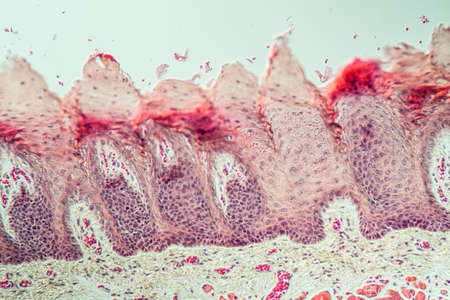
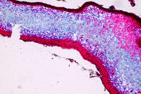
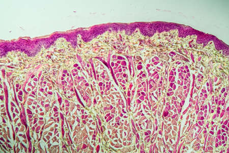
Tongue Tissue with taste buds across 200x
Коллекция по умолчанию
Коллекция по умолчанию
Создать новую
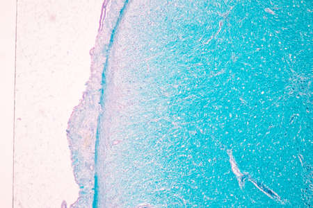
Cerebellum, Thalamus, Medulla oblongata, Spinal cord and Motor Neuron human under the microscope in Lab.
Коллекция по умолчанию
Коллекция по умолчанию
Создать новую
Johannes berry fruit cross 100x
Коллекция по умолчанию
Коллекция по умолчанию
Создать новую

Stomach tissue under the microscope 100x
Коллекция по умолчанию
Коллекция по умолчанию
Создать новую
Tissue of Stomach Human under the microscope in Lab.
Коллекция по умолчанию
Коллекция по умолчанию
Создать новую
Signet ring cell carcinoma of the stomach, light micrograph, photo under microscope
Коллекция по умолчанию
Коллекция по умолчанию
Создать новую
Stomach tissue under the microscope 100x
Коллекция по умолчанию
Коллекция по умолчанию
Создать новую
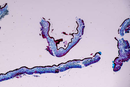
Characteristics of Lichen, hyphae and Symbiotic algae under the microscope for education.
Коллекция по умолчанию
Коллекция по умолчанию
Создать новую
Columnar epithelium of human gall bladder under the microscope in Lab.
Коллекция по умолчанию
Коллекция по умолчанию
Создать новую

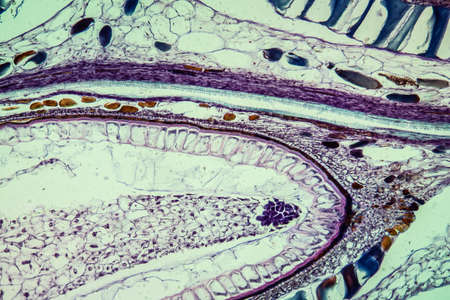
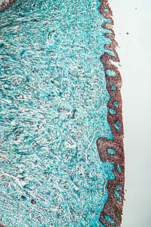
Cell- science background. Esophagus of the dog- cross section
Коллекция по умолчанию
Коллекция по умолчанию
Создать новую
Painting acrylic paint- abstract drawing. Texture background
Коллекция по умолчанию
Коллекция по умолчанию
Создать новую
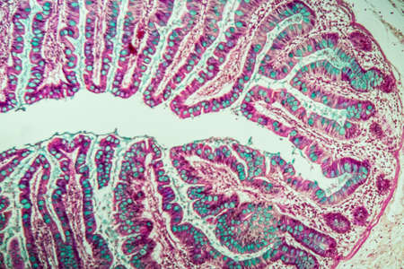
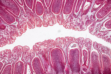
Small intestine with villi under the microscope 100x
Коллекция по умолчанию
Коллекция по умолчанию
Создать новую

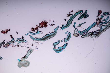
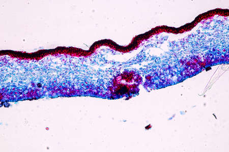
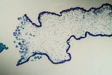
Cross-section through the lichen symbiote body 100x
Коллекция по умолчанию
Коллекция по умолчанию
Создать новую
Tongue Tissue with taste buds across 100x
Коллекция по умолчанию
Коллекция по умолчанию
Создать новую
Intestinal wall section showing villi and epithelial lining clearly
Коллекция по умолчанию
Коллекция по умолчанию
Создать новую

Tissue of Small intestine (Duodenum) and Vermiform appendix Human under the microscope in Lab.
Коллекция по умолчанию
Коллекция по умолчанию
Создать новую
Histopathology of human kidney cross section under microscope for education.
Коллекция по умолчанию
Коллекция по умолчанию
Создать новую
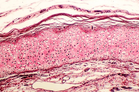
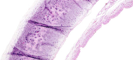
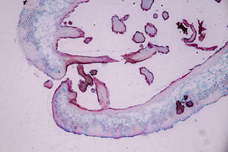
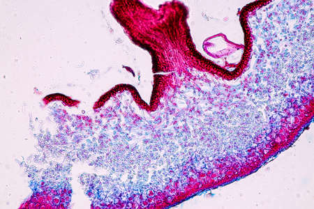
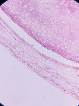
Elastic cartilage of human outer ear, light micrograph
Коллекция по умолчанию
Коллекция по умолчанию
Создать новую
Asthma of the lungs diseased tissue under the microscope 100x
Коллекция по умолчанию
Коллекция по умолчанию
Создать новую

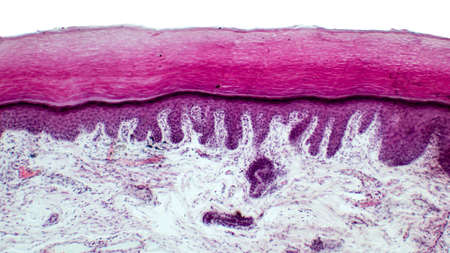
micrograph of medical science stratified squamous epithelium tissue cell
Коллекция по умолчанию
Коллекция по умолчанию
Создать новую

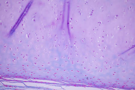
Anatomy and Histological Bone, Elastic cartilage human and Joint of human foetus under the microscope for education.
Коллекция по умолчанию
Коллекция по умолчанию
Создать новую
Characteristics of Lichen, hyphae and Symbiotic algae under the microscope for education.
Коллекция по умолчанию
Коллекция по умолчанию
Создать новую
Cerebellum, Thalamus, Medulla oblongata, Spinal cord and Motor Neuron human under the microscope in Lab.
Коллекция по умолчанию
Коллекция по умолчанию
Создать новую
Breast cancer of the woman diseased tissue 100x
Коллекция по умолчанию
Коллекция по умолчанию
Создать новую
Characteristics of Lichen, hyphae and Symbiotic algae under the microscope for education.
Коллекция по умолчанию
Коллекция по умолчанию
Создать новую
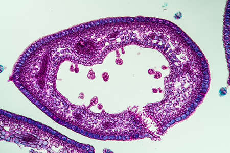
Heather leaf cross section under the microscope, 200x
Коллекция по умолчанию
Коллекция по умолчанию
Создать новую
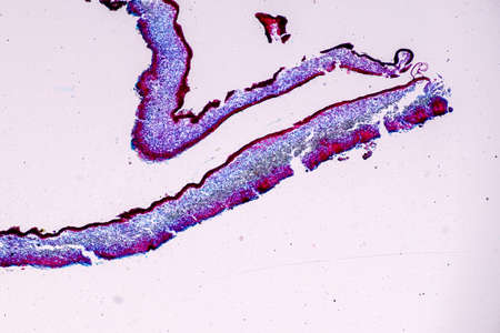
Leech cross section showing internal anatomical structures stained
Коллекция по умолчанию
Коллекция по умолчанию
Создать новую

micrograph of medical science stratified squamous epithelium tissue cell
Коллекция по умолчанию
Коллекция по умолчанию
Создать новую
Pancreas cancer, light micrograph, photo under microscope
Коллекция по умолчанию
Коллекция по умолчанию
Создать новую

Bowen's Disease Tumor under the microscope 100x
Коллекция по умолчанию
Коллекция по умолчанию
Создать новую
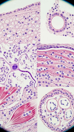
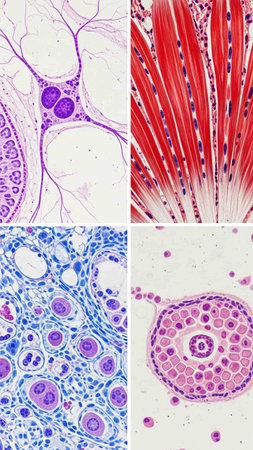
Pathology and Histology Tissue of Mammals under microscope.
Коллекция по умолчанию
Коллекция по умолчанию
Создать новую
Human skin with a scabies infection, Sarcoptes scabiei, under the microscope and with coloration.
Коллекция по умолчанию
Коллекция по умолчанию
Создать новую
Tissue of Small intestine (Duodenum) and Vermiform appendix Human under the microscope in Lab.
Коллекция по умолчанию
Коллекция по умолчанию
Создать новую
Characteristics of Lichen, hyphae and Symbiotic algae under the microscope for education.
Коллекция по умолчанию
Коллекция по умолчанию
Создать новую
Characteristics of Lichen, hyphae and Symbiotic algae under the microscope for education.
Коллекция по умолчанию
Коллекция по умолчанию
Создать новую
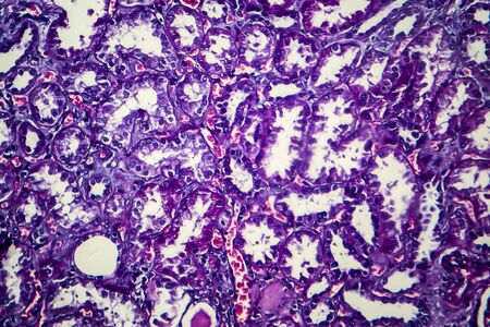
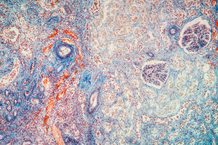
Chronic nephritis, light micrograph, photo under microscope
Коллекция по умолчанию
Коллекция по умолчанию
Создать новую
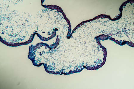
Cross-section through the lichen symbiote body 100x
Коллекция по умолчанию
Коллекция по умолчанию
Создать новую
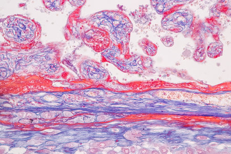
Histological Uterus human, Uterine tube human, Placenta human and Umbilical cord Human under the microscope for education.
Коллекция по умолчанию
Коллекция по умолчанию
Создать новую
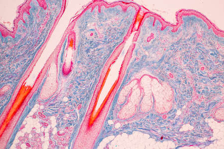
Scalp and hair follicles of human under the microscope in Lab.
Коллекция по умолчанию
Коллекция по умолчанию
Создать новую
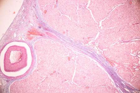
Anatomy and Histological Ovary, Testis and Sperm human cells under microscope.
Коллекция по умолчанию
Коллекция по умолчанию
Создать новую
Histopathology of cirrhosis under the microscope 100x.
Коллекция по умолчанию
Коллекция по умолчанию
Создать новую
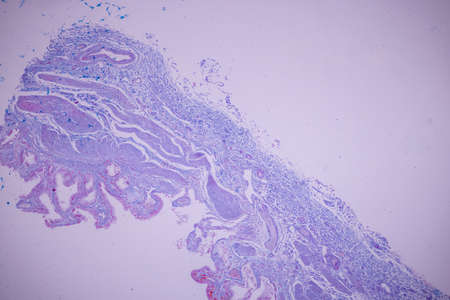
Histology of human tissue, show tracheitis as seen under the microscope
Коллекция по умолчанию
Коллекция по умолчанию
Создать новую
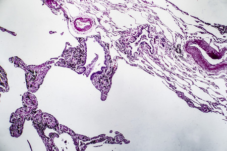
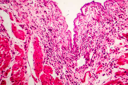
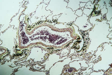
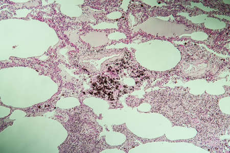
Histopathology of lung emphysema, light micrograph, photo under microscope showing enlargement of air spaces in lung tissue and destruction of alveolar septa
Коллекция по умолчанию
Коллекция по умолчанию
Создать новую
Abstract colorful wet paint
Коллекция по умолчанию
Коллекция по умолчанию
Создать новую
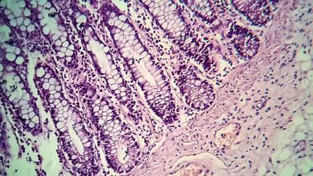
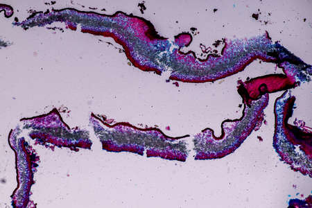
Bacillary dysentery, light micrograph, photo under microscope showing presence of bacteria and accumulation of inflammatory cells in intestinal epithelium
Коллекция по умолчанию
Коллекция по умолчанию
Создать новую

Columnar epithelium of human gall bladder under the microscope in Lab.
Коллекция по умолчанию
Коллекция по умолчанию
Создать новую
Rows of microscope glass slide in the cells
Коллекция по умолчанию
Коллекция по умолчанию
Создать новую
Colon cancer tissue under the microscope 100x
Коллекция по умолчанию
Коллекция по умолчанию
Создать новую
Columnar epithelium of human gall bladder under the microscope in Lab.
Коллекция по умолчанию
Коллекция по умолчанию
Создать новую

Histological Uterus human, Uterine tube human, Placenta human and Umbilical cord Human under the microscope for education.
Коллекция по умолчанию
Коллекция по умолчанию
Создать новую
Basal cell cancer Diseased tissue 100x
Коллекция по умолчанию
Коллекция по умолчанию
Создать новую
Atrophy kidney tissue under the microscope 100x
Коллекция по умолчанию
Коллекция по умолчанию
Создать новую

Cross section of human skin under microscope view for education in laboratory.
Коллекция по умолчанию
Коллекция по умолчанию
Создать новую
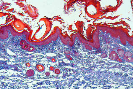
Punch Biopsy of the Skin. Layers of human skin. Hematoxlyn and eosin stain.
Коллекция по умолчанию
Коллекция по умолчанию
Создать новую
Histopathology of human hair follicle, 3D illustration.
Коллекция по умолчанию
Коллекция по умолчанию
Создать новую

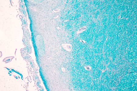
Education anatomy and Histological sample Spinal cord Tissue under the microscope.
Коллекция по умолчанию
Коллекция по умолчанию
Создать новую
Cliated epithelium of human under the microscope in Lab.
Коллекция по умолчанию
Коллекция по умолчанию
Создать новую
Pathology and Histology Tissue of Mammals under microscope.
Коллекция по умолчанию
Коллекция по умолчанию
Создать новую
Leech cross section showing internal anatomical structures stained
Коллекция по умолчанию
Коллекция по умолчанию
Создать новую

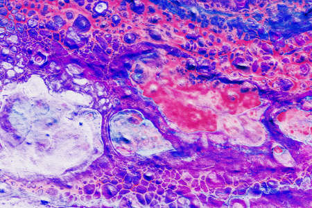
Bladder cat- cell nature background. Abstract- photo macro sections with high magnification with light microscope
Коллекция по умолчанию
Коллекция по умолчанию
Создать новую
Rows of microscope glass slide in the cells
Коллекция по умолчанию
Коллекция по умолчанию
Создать новую
Charcoal dust lung tissue under the microscope 100x
Коллекция по умолчанию
Коллекция по умолчанию
Создать новую
Nasal cavity
Коллекция по умолчанию
Коллекция по умолчанию
Создать новую
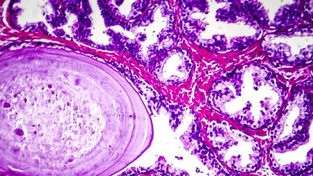
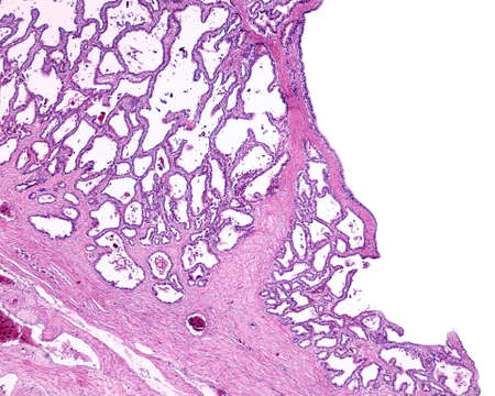
Histopathology of prostate gland hyperplasia, light micrograph, photo under microscope
Коллекция по умолчанию
Коллекция по умолчанию
Создать новую
Characteristics of Lichen, hyphae and Symbiotic algae under the microscope for education.
Коллекция по умолчанию
Коллекция по умолчанию
Создать новую
Columnar epithelium of human gall bladder under the microscope in Lab.
Коллекция по умолчанию
Коллекция по умолчанию
Создать новую

Host cells with spores (mold) are inside wood under the microscope for education.
Коллекция по умолчанию
Коллекция по умолчанию
Создать новую
Characteristics of Lichen, hyphae and Symbiotic algae under the microscope for education.
Коллекция по умолчанию
Коллекция по умолчанию
Создать новую
Human pancreas in the form of a cross section.
Коллекция по умолчанию
Коллекция по умолчанию
Создать новую

Colon polyp, one of the largest polyps
Коллекция по умолчанию
Коллекция по умолчанию
Создать новую
science medical anthropotomy physiology microscopic section of human thyroid gland background
Коллекция по умолчанию
Коллекция по умолчанию
Создать новую

Columnar epithelium of human gall bladder under the microscope in Lab.
Коллекция по умолчанию
Коллекция по умолчанию
Создать новую
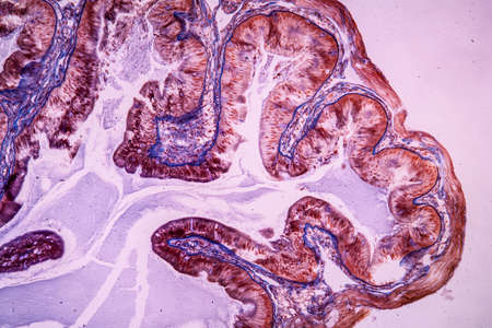
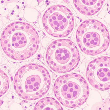
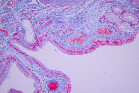
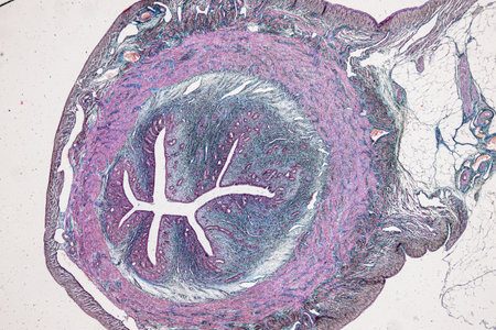
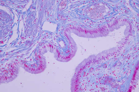
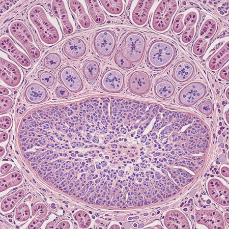
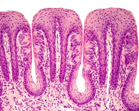
Human seminal vesicle. The surface of the mucosa is very folded. The spaces that look like glands are really infoldings of the mucosa that communicate with the lumen. The epithelium is pseudostratified columnar with basal cells.
Коллекция по умолчанию
Коллекция по умолчанию
Создать новую

Nature pattern: cross-section weevil rye. Beautiful vegetable background. Biological prepared microscope slides
Коллекция по умолчанию
Коллекция по умолчанию
Создать новую
Columnar epithelium of human gall bladder under the microscope in Lab.
Коллекция по умолчанию
Коллекция по умолчанию
Создать новую
Squamous cell carcinoma diseased tissue under the microscope 100x
Коллекция по умолчанию
Коллекция по умолчанию
Создать новую
Characteristics of Lichen, hyphae and Symbiotic algae under the microscope for education.
Коллекция по умолчанию
Коллекция по умолчанию
Создать новую
Tongue Tissue with taste buds across 100x
Коллекция по умолчанию
Коллекция по умолчанию
Создать новую

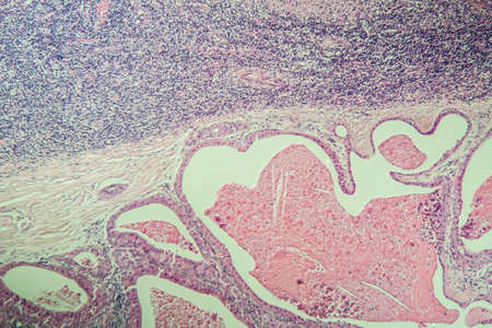
Endometriosis, a disorder in which cells similar to those in the endometrium grow outside the uterus. Light micrograph, photo under microscope
Коллекция по умолчанию
Коллекция по умолчанию
Создать новую
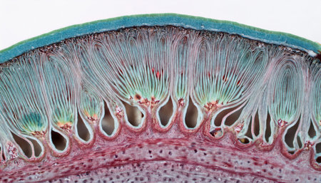
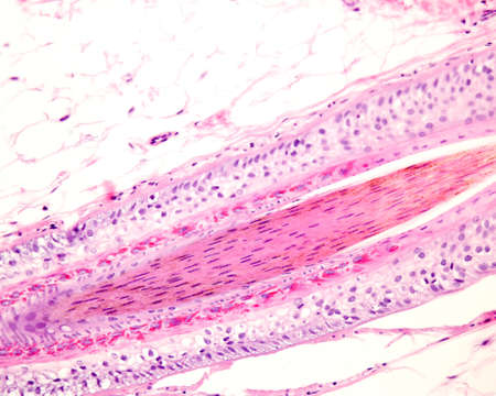
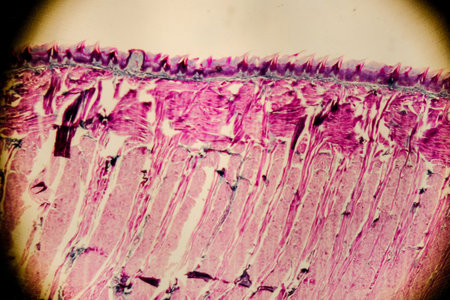
Taste buds in foliate tongue papillae. Many of them show the taste or gustatory pore. Hematoxylin & eosin stain.
Коллекция по умолчанию
Коллекция по умолчанию
Создать новую
Kidney cancer, light micrograph, photo under microscope. High magnification
Коллекция по умолчанию
Коллекция по умолчанию
Создать новую

Histopathology of lung emphysema, light micrograph, photo under microscope showing enlargement of air spaces in lung tissue and destruction of alveolar septa
Коллекция по умолчанию
Коллекция по умолчанию
Создать новую
Cross-section through the lichen symbiote body 100x
Коллекция по умолчанию
Коллекция по умолчанию
Создать новую
This visual representation reveals a cross-section of a plant stem, displaying the intricate structures essential for plant physiology.
Коллекция по умолчанию
Коллекция по умолчанию
Создать новую
Lung tissue as dust lung under the microscope 100x
Коллекция по умолчанию
Коллекция по умолчанию
Создать новую
Acute pyelonephritis, light micrograph, photo under microscope
Коллекция по умолчанию
Коллекция по умолчанию
Создать новую

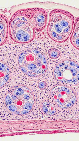
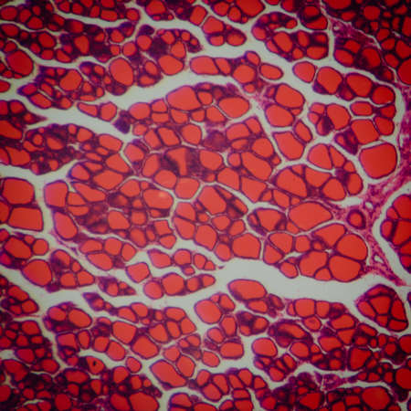
Endemic goiter, light micrograph, abnormal enlargement of the thyroid gland due to dietary iodine deficiency. Photomicrograph shows follicles of varying size, abundant colloid, lymphocytic infiltrate
Коллекция по умолчанию
Коллекция по умолчанию
Создать новую
Salivary gland swollen diseased tissue under the microscope 100x
Коллекция по умолчанию
Коллекция по умолчанию
Создать новую
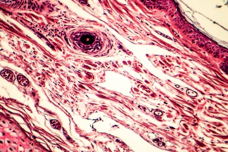
Layers of a human hair follicle in longitudinal section. From outside: hyaline layer, outer root sheath, Henle’s and Huxley’s layers, inner root sheath, and hair shaft in the center.
Коллекция по умолчанию
Коллекция по умолчанию
Создать новую
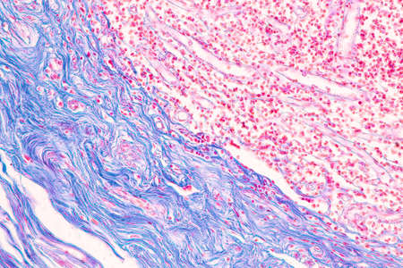
Connective tissue located at human outer ear, light micrograph. Hematoxylin and eosin stain
Коллекция по умолчанию
Коллекция по умолчанию
Создать новую
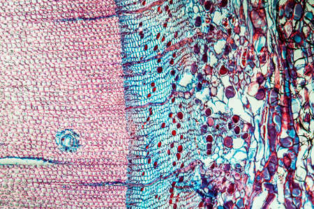
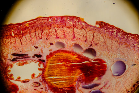
Pine tree trunk across 100x
Коллекция по умолчанию
Коллекция по умолчанию
Создать новую
Histopathology of human under microscope view for education in laboratory.
Коллекция по умолчанию
Коллекция по умолчанию
Создать новую

Host cells with spores (mold) are inside wood under the microscope for education.
Коллекция по умолчанию
Коллекция по умолчанию
Создать новую
Human tongue section showing papillae and stratified epithelium
Коллекция по умолчанию
Коллекция по умолчанию
Создать новую
Abstract bright shiny color fluid background, hand drawn alcohol painting with golden streaks. Liquid ink technique texture for high resolution backdrop design
Коллекция по умолчанию
Коллекция по умолчанию
Создать новую
Legion-Media
Создайте свои проекты на основе качественных стоковых фотографий и видео.


Copyright © Legion-Media.